
Back to the Basics – Airway Anatomy, Part 2

How often do you think about breathing? Probably not very often—unless you find yourself struggling to breathe. In those moments, breathing becomes the only thing you can focus on. This concept carries into this month’s blog as we turn our attention to the lower respiratory tract and review the anatomy that plays a critical role in both normal respiration and respiratory disease.
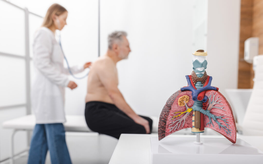
Anatomy of the Intrathoracic Airway
The lower airway includes the trachea, mainstem bronchi, multiple generations of bronchi and bronchioles, alveolar ducts, and alveoli. Each structure contributes uniquely to airflow, gas exchange, and airway protection.
The trachea is often referred to as the “breathing tube.” It is a multilayered structure composed of cartilage and smooth muscle that extends from approximately the sixth or seventh cervical vertebra to the fifth or sixth thoracic vertebra. At birth, the trachea measures about 3 centimeters in length and grows to approximately 10–12 centimeters in adulthood.
The anterior and lateral walls of the trachea are formed by C‑shaped rings of hyaline cartilage, while the posterior wall consists of smooth muscle. This musculature allows the trachea to contract and plays an essential role in effective coughing.
Weakness of the posterior tracheal wall can lead to expiratory central airway collapse (ECAC), a condition that may present with symptoms similar to asthma. ECAC includes both excessive dynamic airway collapse and tracheobronchomalacia (TBM). Excessive dynamic airway collapse occurs due to weakness and atrophy of the posterior tracheal wall, whereas TBM involves abnormal motion of the cartilaginous portion of the trachea.
ECAC may occur in isolation or in combination with other airway diseases, including asthma, chronic obstructive pulmonary disease (COPD), and vocal cord dysfunction. The prevalence of ECAC has been estimated at approximately 13% in the general population and may increase to as high as 37% in individuals with underlying airway disease. Much like asthma, the symptoms of ECAC are common and nonspecific. Pulmonary function tests (PFTs) may demonstrate obstructive or restrictive patterns, making diagnosis challenging. Bronchoscopy remains the most reliable diagnostic tool. Because ECAC can exist with or without asthma—and requires different management—accurate diagnosis is essential.
The Bronchial Tree
Distal to the trachea, the lungs divide into approximately 23 generations of branching airways, collectively known as the bronchial tree. The trachea bifurcates into the right and left main bronchi, which ventilate the lungs.
The right main bronchus is wider, shorter, and more vertical than the left, while the left main bronchus is narrower, longer, and more oblique. Each main bronchus divides into lobar bronchi. The right lung contains three lobes—superior, middle, and inferior—while the left lung contains two lobes, superior and inferior.
The lobes are further divided into segmental bronchi. The right lung has ten segmental bronchi, while the left typically has eight. These airways continue to branch into smaller bronchi and bronchioles, ultimately terminating in the terminal bronchioles, which lead to the alveolar ducts and alveolar sacs. Gas exchange occurs in these most distal regions of the lung.
The lungs are covered by a double‑layered membrane known as the pleura, consisting of the visceral and parietal pleura. The visceral pleura adheres to the lung surface, while the parietal pleura lines the inner chest wall. Between them lies the pleural space, which contains a small amount of pleural fluid. This fluid allows for smooth, frictionless—and therefore painless—movement of the lungs during respiration.
Alveoli and Gas Exchange
Portions of the airway that function solely to conduct air are referred to as dead space, as they do not participate in gas exchange. Even in healthy lungs, approximately one‑third of tidal volume is considered dead space by design.
The alveoli are located at the ends of the bronchioles and are the primary sites of gas exchange. These tiny, grape‑like sacs are microscopic in size but remarkably complex. Millions of alveoli are present in each lung, dramatically increasing the surface area available for oxygen and carbon dioxide exchange. Surrounding capillaries ensure efficient and rapid gas exchange between the air and bloodstream.
By regulating carbon dioxide levels, the alveoli indirectly assist in maintaining normal blood pH. Alveoli are lined with surfactant, a substance that reduces surface tension and prevents collapse during exhalation. In addition to their role in gas exchange, alveoli contribute to immune defense by helping remove pathogens and harmful particles. They also support vocalization by providing the airflow and pressure necessary for sound production. The elastic properties of alveoli help minimize energy expenditure during breathing.
Diseases affecting the alveoli can have significant clinical consequences. Emphysema, for example, destroys alveolar walls and reduces surface area for gas exchange, leading to breathlessness. While emphysema and asthma may share similar symptoms, their pathophysiology, treatment, and prognosis differ substantially. Emphysema is irreversible, whereas asthma can often be well controlled. Importantly, patients may have both conditions simultaneously, typically resulting in more severe symptoms and more frequent exacerbations than either condition alone.
Why the Basics Matter
The respiratory system functions quietly and efficiently—until it does not. When breathing becomes difficult, every detail of the airway matters. Revisiting and deepening our understanding of airway anatomy strengthens clinical judgment, supports accurate diagnosis, and helps differentiate conditions with overlapping symptoms but very different treatments. A solid grasp of the basics ultimately allows clinicians to better support patients when breathing becomes their greatest challenge.
Author:
Heather Murgatroyd, BA, RRT, CPFT, AE-C
Senior Clinical Specialist
Methapharm Respiratory
References